


In case of severe internal diseases, malnutrition and as the age progresses, the growth of the nail slows down and its structure changes. Only a physician can determine the exact cause of the violation based on the results of examinations and microscopic examinations.
However, if you want to get an idea of what is happening to your fingernails or hands, you can use the photo with different types of fungal diseases.
Causes of nail deformity
Molds, yeasts, and dermatophyte fungi cause infectious onychomycosis that shows similar symptoms.
All kinds of nail or hand nail fungus deforms the nail plate, changes its transparency, shine, color, this variety can be seen in the photos presented.
Nail changes occur not only in onychomycosis, but also in injuries, chronic paronychia, psoriasis, hand eczema, dermatitis. Before concluding that you have a fungal infection, you should consider all possible options.
Signs of a fungal infection
The most informative signs of fungal infection are discoloration of the nail plate, the presence of nail detachment, superficial changes - cross-sectional, longitudinal grooves on the nail plate, precise depressions, thickening, nail destruction.
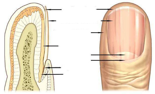
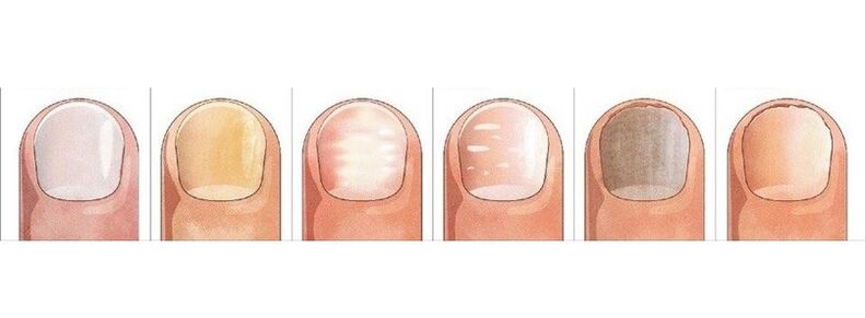
The pink of a healthy nail is determined by the transparency of the nail plate and the blood vessels visible through it. In the case of onychomycosis, the nail loses its transparency, its color becomes brownish, yellow, less often green, black.
Candida fungi and dermatophytes cause onycholysis - the separation of the affected part of the nail. When infected with dermatophytes, onycholysis is observed from the distal edge of the nail, and in Candida infection, the nail lags behind the nail bed at the base, in the area of the crescent.
Candidate fungus may be characterized by inflammation of the lateral periungal spines - paronychia. There are bacterial forms of this disease caused by streptococci and staphylococci as well as non-infectious ones - eczema, psoriasis, systemic vasculitis.
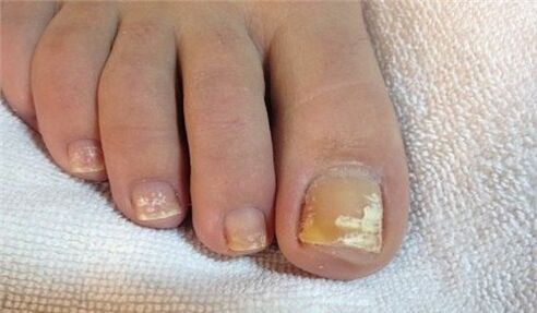
When the toenails are touched by the fungus Trichophyton rubrum, the disc is touched, as shown in the photo, the roller is not affected by the infection. The plate turns yellow, thickens strongly, and the accumulated mushroom masses can be well distinguished underneath.
Nail fungus due to dermatophyte infection
In 95% of all fungal cases, the disease is caused by dermatophytes caused by Trichophyton rubrum and Trichophyton mentagrophytes.
Trichophyton rubrum infection
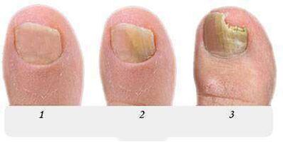
Onichomycosis begins when a fungus penetrates from the free edge under the nail plate. Fungal infection is indicated by a yellowish patch, an uneven, crumbling surface on the distal (distant) edge of the nail in the area of the patch.
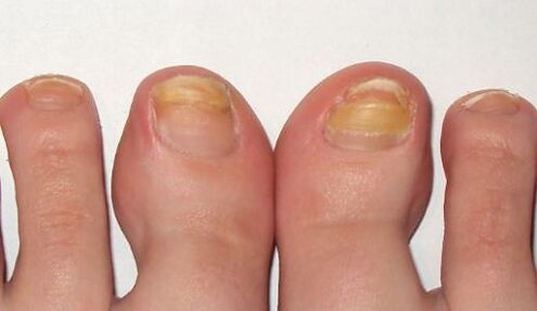
Thedistal-lateral form of the Trichophyton rubrum dermatophyte fungal infectionis common. The photo shows that the spot caused by the introduction of the fungus is located along the lateral periungual nail fold.
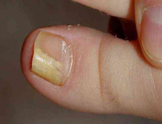
The Trichophyton rubrum fungus usually affects the big toes, causing hyperkeratosis - an accumulation of fungi between the nail plate and the nail bed that appears in the photo as a loose yellowish mass.
At this stage, the fungus occupies an insignificant part of the nail, as in the photo shown, and topical treatment can be used to combat the onset of onychomycosis.
Without treatment, the spot grows, gradually touching the entire edge of the nail and then moving toward the crescent. In the photo, the nail fungus resembles yellowish streaks directed toward the growth zone of the nail plate.
with the distal form of the nail fungus, often found on the big toes, on the distal edge of the nail, in the middle of which a yellowish spot of infection appears, as can be seen in the photo.
In the advanced state of the fungus, more nails are affected on the legs than in the photo, and treatment is no longer limited to topical remedies and pills. In addition to antifungal medications, the nail is subjected to hardware cleaning to partially or completely remove the nail plate.
All known antifungal agents and treatments should be used for long-term therapy of the foot caused by Trichophyton rubrum with hyperkeratosis, as shown in the photo.
A fungal infection with complete nail damage spreads to the entire area of the nail plate, and the nail is completely destroyed.
Another fungal infection of dermatophytes, Trichophyton mentagrophytes, can also lead to a complete fungal infection of the nail.
Trichophyton mentagrophytes infection
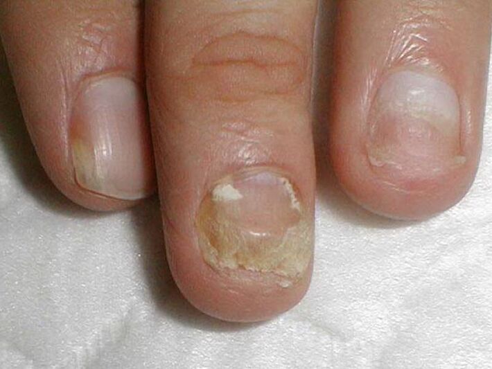
By completely defeating the nail with Trichophyton mentagrophytes fungi, the nail plate is deformed, the photo shows that it thickens, changes its structure, collapses, and yellow spots appear on its entire surface.
Infection of the nail with this dermatophyte usually causes superficial white onicomycosis of the big toe, less often of the little finger.
This fungus is virtually non-existent on the nails of the hand, often causing interdigital dermatophytosis on the feet as in the photo, and requires simultaneous treatment of the skin of the feet and nails.
The symptom of a nail fungus infection, usually on the legs, with white spots of different sizes than in the photo, is reminiscent of leukonychia - a disease of the nail plate.
But unlike leukonychia, in which white spots are caused by air bubbles appearing in the layers of the nail, white spots in fungal infections are the result of the activity of Trichophyton mentagrophytes.
Rarely, superficial white onychomycosis is caused by molds, in AIDS this type of fungus can be caused by Trichophyton rubrum and can affect both the feet and the fingernails.
Nail changes due to Candida infection
The fungus usually occurs in women, affecting the nails of the working hand, which are more likely to come into contact with water.
Candidate onychomycosis is characterized by a proximal form of infection in which the fungus first affects the nail drive at the base of the nail and then penetrates the growth zone and the nail bed. It then gradually moves along the nail from the base to the edge and captures an increasing area of the nail plate.
In candidiasis onychomycosis, the disease is caused by Candida albicans. This fungus attacks the nails and toenails, extending from the crescent zone at the base of the nail plate to the free edge, as shown in the photo.
A sign of Candida nail infection.Candida albicans can penetrate the nail and from its free edge. In this case, they speak of a distal form of infection that is usually combined with candidiasis of the skin.
Treating the candida fungus on the fingernails of the hands and feet, damaging more than half of the area of the nail plate, as in the photo, involves not only fighting onychomycosis, , in the mucous membranes of the genitals. . .
Mold infection
Molds are much less likely to cause fungi than Candida or dermatophytes. The main symptom of nail mold infection, as shown in the photo, is to change the color of the nail plate to blue, black, green in.
Signs of nail mold can be dark spots, dots on the nail plate, or, as in the photo, a black longitudinal stripe.
Antifungal preparations
Antifungal agents with fluconazole, ketoconazole, terbinafine, itraconazole, griseofulvin are used to treat nail fungi caused by dermatophytes, as shown in this photo.
Antifungal agents containing terbinafine are effective in dermatophytic infections.
Antifungals containing voriconazole are very active against dermatophytes.
Thisandare used to treat nail moldon the feet, hands, andto treat candida yeast. The spectrum of action includes molds such as Aspergillum, Fusarium, Penicillium.
Itraconazole-based formulations cope with molds.
Fungal nail diseases
Grayscalesometimes appears on the nail aseczematous. In this case, the nail plate may move away from the nail bed, which may be observed by a fungus.
Externally, themanifestations of psoriasisare very similar. With this disease, not only thediscolorationbut also thenail plate thickens.
There are dot pressures on the surface, note the separation of the nail plate from the nail bed. But there are differences from the fungus: in psoriasis, the detached and healthy parts of the nail are separated over time by a pink, yellowish streak.
Blue colorpseudomonas nail infectiongets nails. Frequent mechanical rubbing of the nail drive causes the appearance of surface grooves, the corrugation of the nail.
White spots in leukonychia, the appearance of which isassociated with metabolic disorders, is also mistaken for a superficial white fungus with a large spot.
Changing the color and shape of your nails will cause injury. Big toes are in the greatest danger. Injured nails as well as a fungus thicken and darken.

The difference between an injury and a fungus is that changes during injury are only detected on the injured finger, the nails of the other fingers remain unchanged, and are not infected with the diseased finger as in onychomycosis.
Trauma can result in the partial separation of the nail from the nail bed, the formation of a cavity that is rapidly planted by the fungi under adverse conditions.
The nail plate can be separated from the nail bed by light (photoonicholysis), iron deficiency anemia, hormonal diseases. Cleavage, loss of nails occurs in cases of lichen erythematosus, bullous dermatosis, nail trauma.
But you can finally make sure the conclusion is right and start treatment, you can only ask for help from a dermatologist - a dermatologist or mycologist - from a doctor treating fungal diseases.